 Quicklinks▼
 myLSUHSC
January Faculty Publications
A new selection of articles have been added to the Faculty Publications display in the Ische Library. These eight articles, as well as all of the articles in our Faculty Publications database, are authored by at least one member of our research community here at LSUHSC-New Orleans. Each month the Library is proud to present copies of eight of these publications in a rotating display of 16.
1. Avegno EM, Middleton JW, Gilpin NW. Synaptic GABAergic transmission in the central amygdala (CeA) of rats depends on slice preparation and recording conditions. Physiol Rep. 2019;7(19):e14245.
2. Conner M, Joshi T, Veerisetty SS, Hutchings J. Coinfection of cytomegalovirus and cryptosporidiosis in a patient with AIDS. ACG Case Rep J. 2019;6(10):e00225.
3. Fontenot J, Spieler B, Hudson C, Boulmay B. Pancreatic acinar cell carcinoma–literature review and case report of a 56-year-old man presenting with abdominal pain. Radiol Case Rep. 2019;15(1):39-43.
4. Harrison-Bernard LM, Naljayan MV, Mercante DE, Gunaldo TP, Edwards S. Longitudinal interprofessional education in a graduate physiology course. Adv Physiol Educ. 2019;43(2):241-245.
5. Hoffman LA, Lufler RS, Brown KM, DeVeau K, DeVaul N, Fatica LM, Mussell J, Byram JN, Dunham SM, Wilson AB. A review of U.S. medical schools’ promotion standards for educational excellence. Teach Learn Med. 2019;1-10.
6. Kerut EK, To F, Summers KL, Sheahan C, Sheahan M. Statistical and machine learning methodology for abdominal aortic aneurysm prediction from ultrasound screenings. Echocardiography. 2019;.
7. Patterson CW, Stalder MW, Richardson W, Steele T, Wise MW, St Hilaire H. Timing of free flaps for traumatic wounds of the lower extremity: Have advances in perioperative care changed the treatment algorithm? J Reconstr Microsurg. 2019;35(8)(8):616-621.
8. Samet JM, Fontham E, Alpirez-Guardao M, Sousa-Santana V. Pollution in the americas: A leading cause of disease burden and an opportunity for cancer pre-vention. Salud Publica Mex. 2019;61(4):417-426.
Publications cited in the Faculty Publications database are harvested weekly from a variety of sources, such as PubMed, SCOPUS, and CINAHL, to name a few. In addition to articles they include books, book chapters, papers, editorials, letters to the editor, and meeting abstracts, all authored by at least one member of the LSUHSC-NO community. The database is maintained by Reference Librarian Kathy Kerdolff and is available to the general public here or via the Library’s webpage. For a PDF of a bibliography of this month’s additions,click here. If you have an article you would like us to highlight or if you have any questions regarding the display or the database, you can contact Kathy Kerdolff.


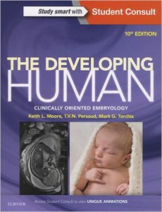 We are pleased to announce that The Developing Human: Clinically Oriented Embryology, 10th edition, is now available in an Ebook format.
 AccessMedicine E-books may be printed one chapter at a time but may not be saved.